DIVISION OF THE NERVOUS SYSTEM
Neuroanatomy: the structure of the nervous system. To learn how the nervous system functions, you must learn how the nervous system is put together.
The nervous system can be divided into several connected systems that function together. Let's start with a simple division:
The nervous system is divided into the
central nervous system and
peripheral nervous system.
Peripheral Nervous System
The peripheral nervous system is divided into two major parts: the somatic nervous system and the autonomic nervous system.
Somatic Nervous System
The somatic nervous system consists of peripheral nerve fibers that send sensory information to the central nervous system AND motor nerve fibers that project to skeletal muscle.
The picture on the left shows the somatic motor system. The cell body is located in either the brain or spinal cord and projects directly to a skeletal muscle.
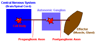
Autonomic Nervous System
 Th
Th
e autonomic nervous system is divided into three parts: the sympathetic nervous system, the parasympathetic nervous system and the enteric nervous system. The autonomic nervous system controls smooth muscle of the viscera (internal organs) and glands.
This picture shows the general organization of the autonomic nervous system. The preganglionic neuron is located in either the brain or the spinal cord. This preganglionic neuron projects to an autonomic ganglion. The postganglionic neuron th
en projects to the target organ. Notice that the somatic nervous system has only one neuron between the central nervous system and the targe
t organ while the autonomic nervous system uses two neurons.

| Divisions of the Nervous System |
 Telencephalon Telencephalon | 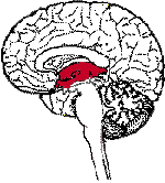 Diencephalon Diencephalon | 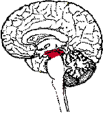 Mesencephalon Mesencephalon |
 Metencephalon Metencephalon | 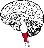 Myelencephalon Myelencephalon |
From a top view, notice how the brain is divided into two halves, called hemispheres. Each hemisphere communicates with the other through the corpus callosum, a bundle of nerve fibers. (Another smaller fiber bundle that connects the two hemispheres is called the anterior commissure).
Some differences between the peripheral nervous system (PNS) and the central nervous system (CNS):
- In the CNS, collections of neurons are called nuclei. In the PNS, collections of neurons are called ganglia.
- In the CNS, collections of axons are called tracts. In the PNS, collections of axons are called nerves.
In the peripheral nervous system, neurons can be functionally divided in three ways:
- Sensory (afferent) - carry information INTO the central nervous system from sense organs or motor (efferent) - carry information away from the central nervous system (for muscle control).
- Cranial - connects the brain with the periphery or spinal - connects the spinal cord with the periphery.
- Somatic - connects the skin or muscle with the central nervous system or visceral - connects the internal organs with the central nervous system.
BRAIN STRUCTURES
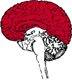
Cerebral Cortex
Functions:
- Thought
- Voluntary movement
- Language
- Reasoning
- Perception
The word "cortex" comes from the Latin word for "bark" (of a tree). This is because the cortex is a sheet of tissue that makes up the outer layer of the brain.
Cerebellum
Functions:
The word "cerebellum" comes from the Latin word for "little brain." The
cerebellum is located behind the brain stem.
Brain stem
Functions: 
- Breathing
- Heart Rate
- Blood Pressure
The brain stem is a general term for the area of the brain between the thalamus and spinal cord.
Hypothalamus
Functions: 
- Body Temperature
- Emotions
- Hunger
- Thirst
- Circadian Rhythms
The hypothalamus is composed of several different areas and is located at the base of the brain. Although it is the size of only a pea (about 1/300 of the total brain weight), the hypothalamus is responsible for some very important functions.
Thalamus
Functions: 
- Sensory processing
- Movement
The thalamus receives sensory information and relays this information to the cerebral cortex.
Limbic System
Functions:
The limbic system (or the limbic areas) is a group of structures that includes the amygdala, the hippocampus, mammillary bodies and cingulate gyrus.
Hippocampus
Functions:
The hippocampus is one part of the limbic system that is important for memory and learning.
Basal Ganglia
Functions:
The basal ganglia are a group of structures, including the globus pallidus, caudate nucleus, subthalamic nucleus, putamen and substantia nigra, that are important in coordinating movement.
Midbrain
Functions:
- Vision
- Audition
- Eye Movement
- Body Movement
The midbrain includes structures such as the superior and inferior colliculi and red nucleus. There are several other areas also in the midbrain.


.jpg)
